40 diagram of a human cell with labels
Diagram of human skin structure — Science Learning Hub Diagram of human skin structure. Add to collection. + Create new collection. Tweet. Rights: University of Waikato Published 1 February 2011 Size: 100 KB Referencing Hub media. The epidermis is a tough coating formed from overlapping layers of dead skin cells. Labeled Plant Cell With Diagrams - Science Trends The parts of a plant cell include the cell wall, the cell membrane, the cytoskeleton or cytoplasm, the nucleus, the Golgi body, the mitochondria, the peroxisome's, the vacuoles, ribosomes, and the endoplasmic reticulum. Parts Of A Plant Cell The Cell Wall Let's start from the outside and work our way inwards.
Animal Cell Diagram with Label and Explanation: Cell ... - Collegedunia Below is the diagram of the animal cell which shows the organelles present in it. The cell is covered with cytoplasm which consists of cell organelles in it. The nucleus is covered with a rough Endoplasmic Reticulum and other organelles each designed for a specific purpose.
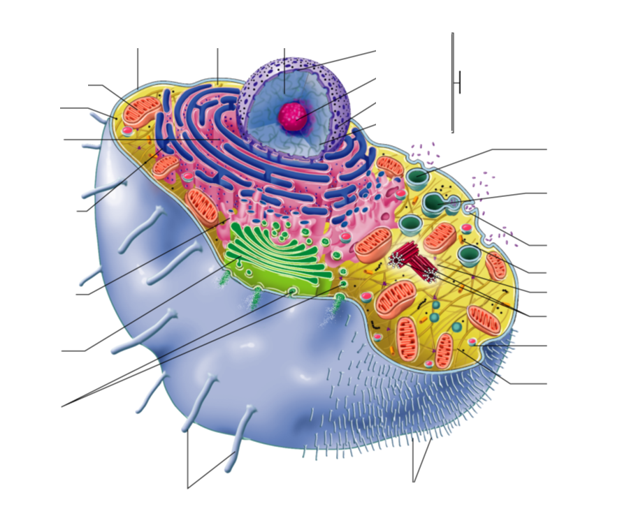
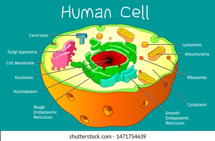
Diagram of a human cell with labels
› books › NBK144066Authentication of Human Cell Lines by STR DNA Profiling ... May 01, 2013 · Since 1951 when the first human cell line, HeLa, was established there has been an increase in the use of human cell lines as models for human diseases such as cancer, substrates for the production of viruses for vaccine production and as tools for the production of recombinant proteins for therapeutics. Unfortunately this accelerated use of human cell lines and the lack of best practices in ... The following diagram shows cells of onion peel label class ... - Vedantu The following diagram shows human cheek cells, label the parts as observed by you. Answer Verified 106.2k + views Hint: The diagrams mentioned above are the internal structure of an onion peel and human cheek cells. In order to label them, we need to understand its anatomy and know about various structures present in it. The Human Skeleton: All You Need to Know - Bodytomy Labeled Skeleton Diagram This skeleton diagram will help explain the different bones of the human body clearly. Cranium The cranium is a skull bone that covers the brain, as seen in the skeleton diagram. The facial bones are not a part of the cranium. The bones that are just above the ear or in front of the ear are known as temporal bones. Stapes
Diagram of a human cell with labels. Karyotype Diagram - SmartDraw Karyotype. A karyotype is a complete set of all chromosomes of a cell of any living organism. Karyotypes are examined in searches for chromosomal aberrations such as genetic disorders, and can also be used to determine other macroscopically visible aspects such as gender. Normal Male Karyotype. Normal Female Karyotype. Human Anatomy Label Me! Printouts - EnchantedLearning.com Human Anatomy Label Me! Elementary-level Printouts. Read the definitions then label the diagrams. Advertisement. EnchantedLearning.com is a user-supported site. As a bonus, site members have access to a banner-ad-free version of the site, with print-friendly pages. ... Animal Cell Anatomy Label the animal cell diagram using the glossary of ... Skeletal System - Labeled Diagrams of the Human Skeleton The skeletal system's cell matrix acts as our calcium bank by storing and releasing calcium ions into the blood as needed. Proper levels of calcium ions in the blood are essential to the proper function of the nervous and muscular systems. Bone cells also release osteocalcin, a hormone that helps regulate blood sugar and fat deposition. PDF Human Cell Diagram, Parts, Pictures, Structure and Functions Diagram of the human cell illustrating the different parts of the cell. Cell Membrane The cell membraneis the outer coating of the cell and contains the cytoplasm, substances within it and the organelle. It is a double-layered membrane composed of proteins and lipids.
› articles › s41592/022/01412-7A pan-tissue DNA methylation atlas enables in silico ... Mar 11, 2022 · For most human tissues and organs, generating DNAm reference profiles for all underlying cell types is very challenging owing to incomplete knowledge of tissue composition and cell-type-specific ... Human Cell Organelles Labeling Diagram - Quizlet Human Cell Organelles Labeling STUDY Learn Flashcards Write Spell Test PLAY Match Gravity Created by Mackenna_Rios5 Terms in this set (8) Vesicles Transports molecules between organelles and the cell membrane Ribosome Makes Protein Mitochondria Makes ATP Smooth ER Makes lipids and vesicles Lysosomes Draw a diagram of typical cell and label the following parts ... Draw a diagram of typical cell and label the following parts in it. Cell membrane. Vacuole Nucleus Endoplasmic reticulum. Mitochondria Golgi body.1 answer · Top answer: Answr has image solution available for this question Animal Cell Diagram Stock Photos and Images - Alamy Animal Cell Anatomy Diagram Structure with all parts nucleus smooth rough endoplasmic reticulum cytoplasm golgi apparatus mitochondria membrane centro. ID: M1X7G4 (RF) Diagram showing anatomy of animal cell illustration. ID: KE1PWP (RF) Animal cell and fungal (yeast) cell structure. cross section and anatomy of cell. Biology Chart.
Circulatory System Labeled Diagram stock illustrations Browse 154 circulatory system labeled diagram stock illustrations and vector graphics available royalty-free, or start a new search to explore more great stock images and vector art. Newest results Heart Poster Heart blood flow circulation anatomical diagram with atrium and... Anatomy of Nerves of Body and Head Bacteria in Microbiology - shapes, structure and diagram Bacteria cells are the smallest living cells that are known; even though viruses are smaller than bacteria, viruses are not living cells. In microbiology there are different types of bacteria with various sizes, shapes, and structures. The bacteria shapes, structure, and labeled diagrams are discussed below. Cell: Structure and Functions (With Diagram) - Biology Discussion Eukaryotic Cells: 1. Eukaryotes are sophisticated cells with a well defined nucleus and cell organelles. 2. The cells are comparatively larger in size (10-100 μm). 3. Unicellular to multicellular in nature and evolved ~1 billion years ago. 4. The cell membrane is semipermeable and flexible. 5. These cells reproduce both asexually and sexually. A Well-labelled Diagram Of Animal Cell With Explanation Animal cells are eukaryotic cells that contain a membrane-bound nucleus. They are different from plant cells in that they do contain cell walls and chloroplast. The animal cell diagram is widely asked in Class 10 and 12 examinations and is beneficial to understand the structure and functions of an animal. A brief explanation of the different ...
Cell Membrane Diagram Labeled : Functions and Diagram Cell Membrane Diagram. There are no organelles in the prokaryotic cells, i.e., they have no internal membrane systems. While lipids help to give membranes their flexibility, proteins monitor and maintain. We all keep in mind that the human body is very elaborate and a technique I found out to understand it is by means of the manner of human ...
› photos › diagram-of-bodyDiagram Of Body Organs Female Pics Pictures, Images ... - iStock Human anatomy scientific illustrations: female reproductive organ Human anatomy scientific illustrations with latin/italian labels: female reproductive organ diagram of body organs female pics stock illustrations
7 Best Images of Neuron Label Worksheet - Blank Neuron Cell Diagram, Synapse Neuron Worksheet ...
Anatomy (Human Body) Labeling - Exploring Nature Arteries of the Lower Limb (Pelvis, Leg and Foot) Labeling. Arteries of the Upper Limb (Shoulder, Arm, Hand) Labeling. Blood Vessel Anatomy Labeling

Questions And Answers On Labeled/Unlebled Diagrams Of A Human Cell / Question 14: Draw a labeled ...
› photos › muscular-systemMuscular System Labeled Diagram Stock Photos, Pictures ... Labeled Muscles of the Human Body, Anterior View, 3D Rendering Frontal view of the muscular system of the male human body with descriptive labels pointing to the muscles on a white background. muscular system labeled diagram stock pictures, royalty-free photos & images
BYJUS BYJUS

Cell Review Guide Answers in 2020 | Human cell structure, Human cell diagram, Animal cell structure
Label Diagram Human Body Stock Illustrations - Dreamstime Download 188 Label Diagram Human Body Stock Illustrations, Vectors & Clipart for FREE or amazingly low rates! New users enjoy 60% OFF. 187,764,331 stock photos online. ... Labeled diagram of the neuron. Nerve cell that is the main part of the nervous system. The circulatory system. The circulatory or cardiovascular human body system medical ...
Label Cell Parts | Plant & Animal Cell Activity | StoryboardThat Click "Start Assignment". Find diagrams of a plant and an animal cell in the Science tab. Using arrows and Textables, label each part of the cell and describe its function. Color the text boxes to group them into organelles found in only animal cells, organelles found in only plant cells, and organelles found in both cell types.
parts of a human cell | Diagram of the human cell illustrating the ... Apr 2, 2014 - parts of a human cell | Diagram of the human cell illustrating the different parts of the cell ... Pinterest. Today. Explore. ... This diagram of a human skeleton is labeled with 12 major bones, from skull to fibula. Nicole. Science. Human Cell Structure. Animal Cell Structure. Plant And Animal Cells.
quizlet.com › 515111566 › ch-8-mastering-biologych 8 mastering biology Flashcards | Quizlet The Human Life Cycle 3 of 16 Review Watch this video and then answer the questions. Part A The figure shows the human life cycle. Can you identify the structures and processes? Drag the labels to their appropriate locations on the figure. Pink labels represent structures, and blue labels represent processes.
What Is Going On Inside That Cell? | Human cell diagram, Cell diagram ... Use the word bank below to identify the parts of the human cell. Did you know?The cell is the basic unit of the human body. There are over one billion cells in each human body. Cells group together to make skin, bones, and blood. Inside the cell nucleus is DNA, which identifies the color of hair, eyes, and skin. DNA also affects the way we look ...
Human Heart Diagram Labeled | Science Trends Daniel NelsonPRO INVESTOR. The human heart is an organ responsible for pumping blood through the body, moving the blood (which carries valuable oxygen) to all the tissues in the body. Without the heart, the tissues couldn't get the oxygen they need and would die. Along with lymphatic vessels, the blood, blood vessels, and lymph, the heart ...
byjus.com › biology › liver-diagramLiver Diagram with Detailed Illustrations and Clear Labels The liver is one of the most important organs in the human body. Anatomically, the liver is a meaty organ that consists of two large sections called the right and the left lobe. The rib cage partly protects the liver and cannot be felt if you were to touch it. However, it can be felt ascending and descending if you were to take a deep breath.
A Labeled Diagram of the Animal Cell and its Organelles A Labeled Diagram of the Animal Cell and its Organelles There are two types of cells - Prokaryotic and Eucaryotic. Eukaryotic cells are larger, more complex, and have evolved more recently than prokaryotes. Where, prokaryotes are just bacteria and archaea, eukaryotes are literally everything else.

The diagram shows a certain kind of cell with all of its major parts labeled. Which statement is ...
sciencequiz.net › newjcscience › jcbiologyThe Cell - ScienceQuiz.net The diagram shows a plant cell as seen under a microscope. Two of the labels are incorrect. What are they? ... A typical human cell has a diameter of 0.00002 metre ...
Human Body Organs Diagram Stock Photos and Images - Alamy Body meridians - Schematic diagram with main acupuncture meridians and their directions of flow. Lungs, heart, liver, stomach, tooth human body organ icon set, flat internal organs silhouette vector symbol collection illustration. Medical diagram showing of the mechanisms of appetite regulation.
Human Cells Printables and Diagrams - The Successful Homeschool These cells include: leukocytes, haematids, thrombocytes, ovum, sperm, sarcomeres, enterocytes, neurons, osteocytes, hepatocytes. They will learn the parts of a cell thanks to a labeled diagram. They will get to see what blood looks like under a microscope without needing to own a microscope. They get to color a cell and then label the parts.

Questions And Answers On Labeled/Unlebled Diagrams Of A Human Cell : Plant Cell Definition ...
Human Cell Diagram, Parts, Pictures, Structure and Functions Diagram of the human cell illustrating the different parts of the cell. Cell Membrane. The cell membrane is the outer coating of the cell and contains the cytoplasm, substances within it and the organelle. It is a double-layered membrane composed of proteins and lipids. The lipid molecules on the outer and inner part (lipid bilayer) allow it to ...


Post a Comment for "40 diagram of a human cell with labels"